PLANVERI
submenu
PLANVERI – system for independent verification of dose calculation in radiotherapy
PLANVERI performs an independent check and verification of the primary planning system's dose calculation algorithm for each patient plan. It uses data in DICOM format.
The necessary input data are:
- CT images in DICOM format (CT),
- contours in DICOM format (structure set, RS),
- treatment plan in DICOM format (RP) and
- dose distribution in DICOM format (RD).
The entire system is fully automatic, there is no need for the operator to transfer or enter data manually.
Data that are not exported from the primary planning system into DICOM (CT, RS, RP, RD) are not processed by the PLANVERI system (possible exceptions must be consulted with the manufacturer).
PLANVERI WEBAPP
The PLANVERI system includes the web application PLANVERI WEBAPP, which provides the user with an overview of the performed calculations, analysis of the results, the current calculation queue and other information.
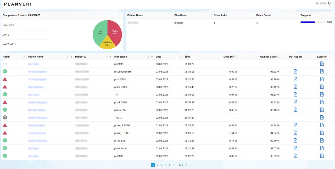
The home page of a web application consists of three main components. In the upper left corner, there is a section that shows users information about the number and results of calculations that were performed during the current day.
In the upper right corner, the status of the calculations that are currently in progress is displayed. Basic information about ongoing calculations and their progress can be seen here.
The rest of the home page is taken up by the plans table, where users are shown key information about each plan, including the patient's name, calculation result, and plan name. On the right side of the table there is a column with options to download a PDF report and open a log of the data processing.
Clicking on a patient's name will open a plan detail allowing for deeper analysis and visualization. In the header of the table columns there are buttons for filtering and changing the order of the results in the table. Only some records from the total number are displayed in the table. To browse between individual pages of records, you can use the navigation below the table.
Symbol
![]()
![]()
![]()
![]()
Status
OK
Failed
Waiting
N/A
Meaning
The dose distribution matching criteria between the two plans were met.
The compared plans differ in some of the parameters by more than the specified matching criteria.
This is a temporary status. This plan is queued for calculation or is currently being calculated.
The plan was not calculated.
Plan detail
The Plan detail opens after clicking on the patient's name in the plan table and is divided into 4 segments. The first section, General Information, contains basic information about the patient and the plan.
It is about:
- final result,
- treatment plan name,
- calculation date,
- computational grid size,
- reference grid size (i.e. the calculation grid of the primary planning system),
- the uncertainty of the Monte Carlo calculation,
- dose depositon method (dose to medium or dose to water), and
- information about the calculation algorithm.

The second part, Result of Plan Comparison, contains a comparison of dose distributions. It is about point dose comparison (isocenter and user-specified point) and structure dose comparison (user-specified structure).

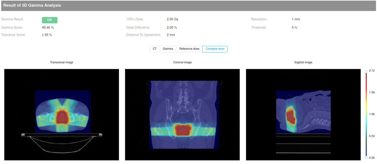
The third part, Result of 3D Gamma Analysis, contains slices of CT images with gamma layer, reference dose distribution or dose distribution calculated in PLANVERI.
When the plan detail is opened, a CT image is displayed overlaid with a gamma layer. Above the images there are buttons for switching the displayed layers. After clicking the "CT" button, only CT sections are displayed, the "Gamma" button adds a gamma layer to the images, other buttons allow the display of reference and comparative doses. Information on the overall 3D gamma result, 3D gamma score, tolerance level, gamma criteria and resolution of the 3D gamma matrix is also provided here.
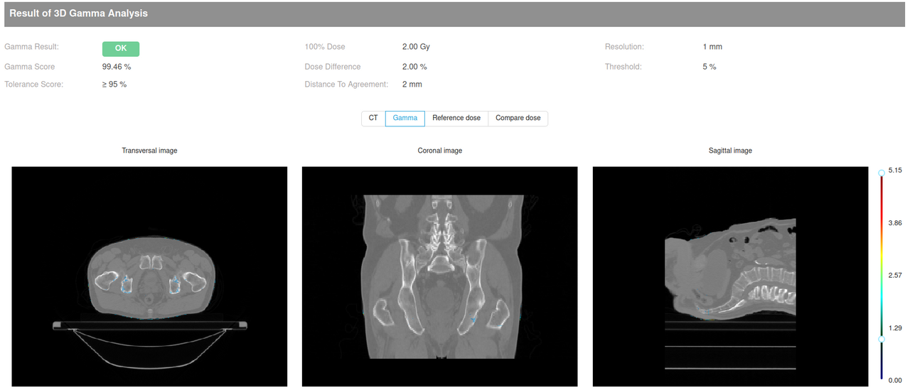 To the right of the images, there is a slider that can be used to set the range of values displayed as a second layer above the CT image itself. This slider allows the user to select a specific range of values to display.
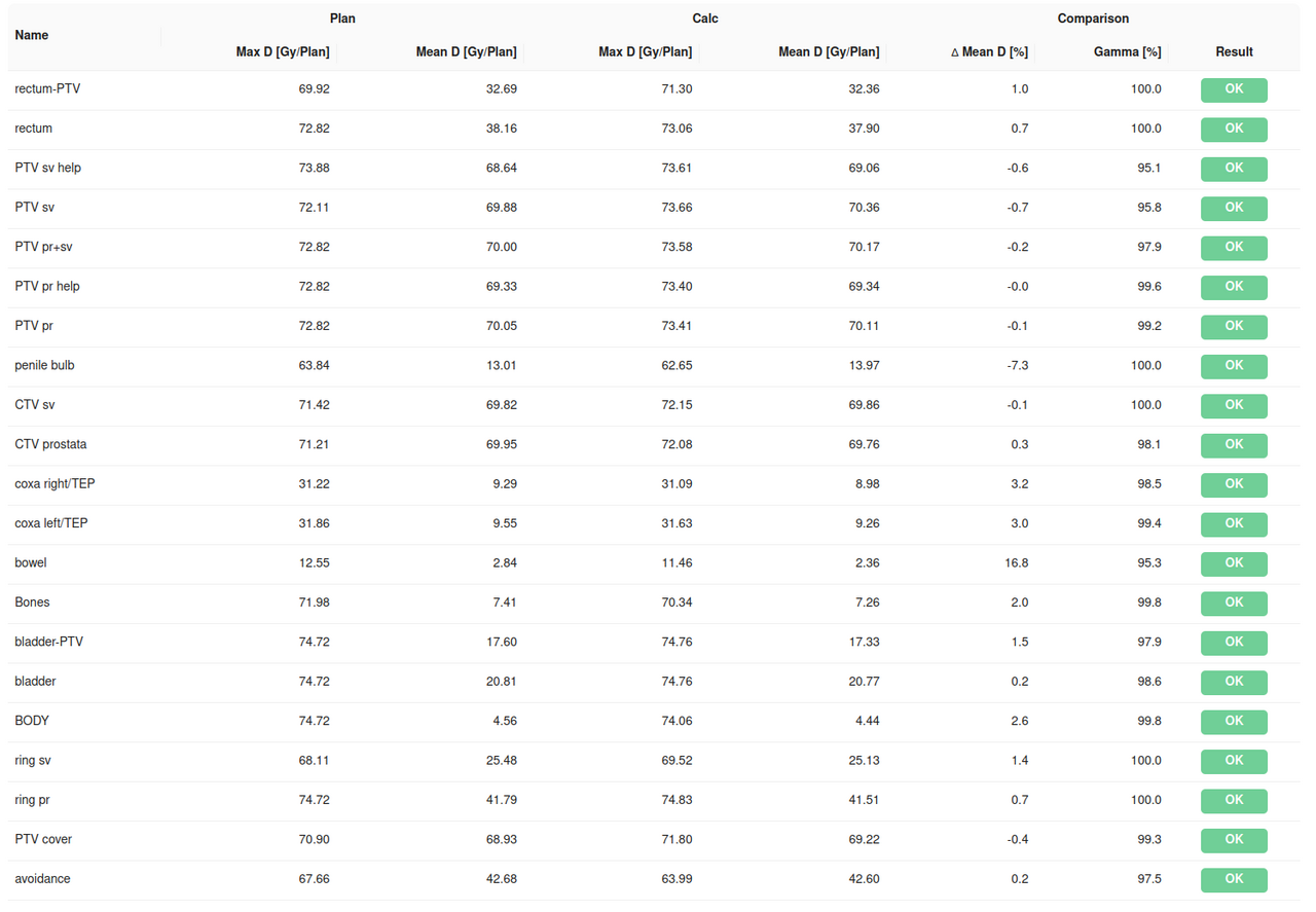
The fourth section of the plan detail, Results of DVH Comparison, contains a graph and tables showing the differences between the planned dose and the calculated dose.
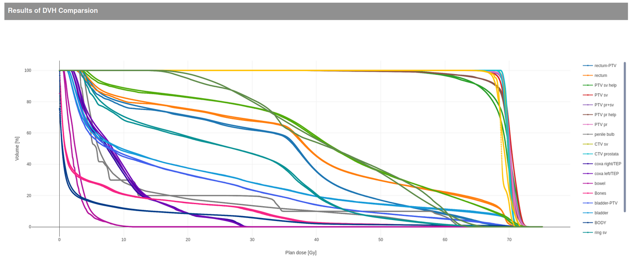 To the right of the graph there is a legend, which is used for quick orientation in the visualized data. Each element in the graph is represented in the legend by a certain color along with a description. Clicking on an item in the legend will hide or show the corresponding element in the chart. Double-clicking on an item in the legend will isolate the given element and hide all others.
Display of information: When the user moves the mouse cursor over a specific point in the graph, a so-called tooltip is displayed. It provides detailed information about the selected point.
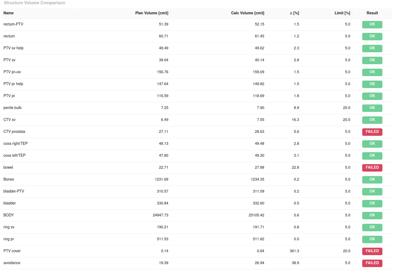
Further, a comparison of dose statistics for individual structures is presented in the form of tables. This is a comparison of mean dose, maximum dose and structure volume.
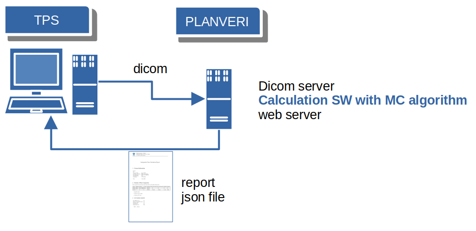
The Process Of Data Processing
The process takes place in the following steps:
- DICOM data is exported from the primary planning system to the PLANVERI calculation server (the export is initiated by the operator).
- if access to the local PACS (or planning system database) is provided, PLANVERI can automatically select DICOM files for calculation
- the system automatically starts calculating the dose distribution for the given radiation plan after the data transfer is complete
- after the calculation is performed, the predefined parameters of the calculated dose distribution and the dose distribution exported from the primary planning system are automatically compared
- the results are accessible via an interactive web interface from any PC located on the same network or by other means.
The data processing process is shown schematically in the following figure
SUPPORTED TREATMENT MACHINE MODELS
Monte Carlo calculation is supported for:
- all types of conventional linear accelerators from manufacturers: Elekta®, Varian® (including HalcyonTM, EthosTM), Siemens
- Accuray® Cyber Knife® (cones, Iris, Incise2), TomoTherapy®, Radixact®
- ZAP-X®

